 By Dr Steven Hewitt — Chiropractor · AHPRA: CHI0001115420 · 28 March 2026
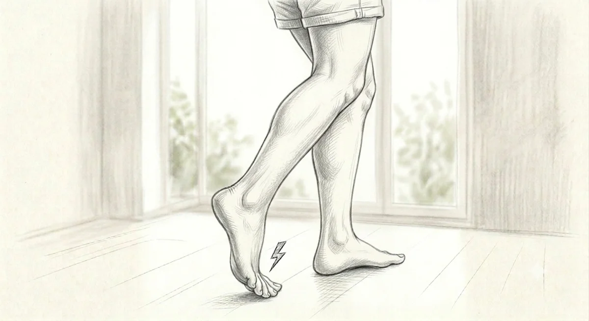
Category: Mechanism Post · Related: Plantar Fasciopathy · Achilles Tendinopathy · Tibialis Posterior Tendinopathy · PFPS · ITB Syndrome
Most people who present with knee pain think about their knee. Most people with persistent plantar fascia pain think about their heel. The research on functional hallux limitus suggests the first place worth looking is sometimes the big toe — specifically, whether it can extend freely during the propulsive phase of gait.
This is not a fringe idea. It is a well-described biomechanical mechanism with a clear anatomical basis, a documented set of compensatory consequences, and a direct connection to several of the most common lower limb conditions we see in clinical practice. It is also one of the most frequently unassessed findings in people who are told they have chronic knee, hip, or plantar fascia problems.
The Windlass Mechanism: Why the Big Toe Matters
The plantar fascia is not a passive structure. It functions as the tension element in the arch — part of a truss-and-beam system that converts the foot into a rigid lever during the propulsive phase of gait. As the heel rises and the body's weight shifts forward over the metatarsal heads, the hallux (big toe) dorsiflexes against the ground. This dorsiflexion winds the plantar fascia tightly around the first metatarsal head — shortening it, increasing tension in the arch, and raising the medial longitudinal arch in preparation for push-off.
This is the windlass mechanism. It is why the foot supinates and becomes rigid at exactly the right moment during gait — not through muscular effort, but through the mechanical geometry of the plantar fascia and first metatarsophalangeal (MTP) joint working together. [1]
For the windlass to function correctly, the first MTP joint needs to achieve at least 60° of passive dorsiflexion during the stance phase. [1] In a healthy foot, this happens automatically with every step. In functional hallux limitus (FHL), it does not.
What Is Functional Hallux Limitus?
Functional hallux limitus is a condition in which the first MTP joint has adequate range of motion in non-weight-bearing — the toe moves freely when the foot is unloaded — but dorsiflexion is blocked or severely restricted during weight-bearing in the closed kinetic chain. [1]
This is the critical distinction. Examining the hallux with the patient lying supine and moving the toe passively will appear normal. The restriction only manifests when the foot is loaded and the first metatarsal is required to plantarflex as the heel rises — precisely when the windlass mechanism must engage.
When it cannot, the first MTP joint shifts from a gliding contact pattern — the normal, energy-efficient geometry of joint loading — to a rolling contact pattern. [1] This increases compressive force on the dorsal joint surfaces, stresses the plantar fascia under abnormal load, and — because the system cannot complete its propulsive function — forces the rest of the kinetic chain to compensate.
Those compensations are where the problems downstream of the foot are produced.
What the Chain Does When the Big Toe Cannot Move
A study by Hall and Nester examined sagittal plane kinematics at the ankle, knee, and hip when first MTP dorsiflexion was artificially restricted in twenty people with symptom-free feet. Even in people with no existing pathology, blocking hallux dorsiflexion with a rigid insole produced measurable changes throughout the chain: the ankle became more dorsiflexed in late midstance, the knee became more flexed during midstance, and the hip became less extended during late midstance. [2]
These are not small, clinically irrelevant changes. They represent the body's attempt to maintain forward progression of the centre of mass when the normal propulsive mechanism is unavailable. The compensation is distributed — a little more ankle, a little more knee, a little less hip — and when repeated thousands of times per day across years, it imposes non-physiological loading on each of those joints.
The rotational consequences are equally significant. A study by Lafuente and colleagues compared the capacity for internal rotation of the lower limb in 45 people with mild hallux limitus versus 35 controls. The hallux limitus group had significantly less lower limb internal rotation capacity (p < .0001), with a strong correlation between hallux dorsiflexion and internal rotational pattern (Spearman r = 0.638; p < .0001). [3]
This matters clinically because internal tibial rotation during the loading phase of gait is the mechanism through which foot pronation drives knee valgus and patellofemoral stress. A foot that cannot complete the windlass mechanism and return to supination at push-off — because the hallux cannot extend — is a foot that delays or reduces the supination signal that normally terminates internal tibial rotation. The result is a lower limb that spends more time in internal rotation under load, placing greater demand on the lateral hip structures (gluteus medius, TFL, iliotibial band) and increasing patellofemoral compression.
This is how a big toe problem becomes a knee problem.
The Plantar Fascia Connection
The relationship between FHL and plantar fasciopathy is bidirectional — and understanding the direction matters for treatment.
A case-control study by Aranda and Munuera compared hallux dorsiflexion in 50 patients with plantar fasciitis against 50 matched controls. The plantar fasciitis group had significantly less hallux dorsiflexion (p < .001), and hallux dorsiflexion was inversely correlated with foot pronation (Spearman −0.441; p < .01). [4] Restricted hallux dorsiflexion was the most consistent finding separating people with plantar fasciitis from those without it.
A cadaveric biomechanical study by Viehöfer and colleagues demonstrated the mechanical mechanism: directly increasing tension in the plantar fascia produced a measurable, significant decrease in hallux extension in all specimens (p < .001), with a maximum mean reduction of 4.2° at the highest tension levels. [5]
This creates the vicious cycle. A tight or irritated plantar fascia mechanically limits hallux dorsiflexion. Limited hallux dorsiflexion impairs the windlass mechanism, increases tensile load on the plantar fascia at push-off, and delays the arch-stiffening that should reduce that load. The two conditions maintain each other — which is why treating only the heel insertion of the plantar fascia while ignoring first MTP mobility so often produces incomplete results.
The Achilles Connection
The upstream driver of FHL in most people is not the first MTP joint itself — it is the posterior chain.
Maceira and Monteagudo describe what they term the Achilles-calcaneus-plantar system: the gastrocnemius-soleus complex, Achilles tendon, calcaneus, and plantar aponeurosis function as a continuous tension system. When the gastrocnemius is restricted — even subclinically, below the threshold of a clinically obvious equinus — it increases tension through the Achilles to the calcaneus, which transfers into increased tension in the plantar aponeurosis. This increased plantar aponeurosis tension reduces the capacity for hallux extension during the stance phase, producing the functional restriction at the first MTP joint. [1]
This has direct implications for how FHL is assessed and managed. Addressing FHL only at the foot — mobilising the first MTP joint, working the sesamoids — without assessing and treating gastrocnemius restriction will address the local joint mechanics while leaving the primary mechanical driver intact. In our clinical practice, assessment of first MTP mobility is always paired with assessment of the gastrocnemius, soleus, and posterior chain tension — because that is where the upstream driver is most commonly found.
The Fascial Angle
The plantar fascia is not just a passive tie-rod — it is a sensorimotor structure embedded in a continuous fascial sleeve that connects the foot to the lower leg, thigh, and ultimately the thoracolumbar fascial system via the posterior myofascial chain. As detailed in anatomical research by Stecco and colleagues, the plantar fascia is continuous with the deep fascia of the leg, blending proximally with the crural fascia. [6]
Densification of the plantar fascial compartment — the hyaluronan-mediated viscosity change that produces stiffness in fascial tissue throughout the body — restricts the mechanical freedom of the plantar fascia to lengthen and shorten as the arch loads and unloads during gait. When the tissue cannot move freely, the windlass mechanism is impaired before first MTP joint restriction even becomes the primary factor.
Manual therapy directed at the plantar fascial compartment and the posterior crural fascia — restoring compliance and glide — addresses the mechanical environment in which the first MTP joint operates. This is distinct from, and complementary to, direct mobilisation of the first MTP joint itself. In our clinical experience, both components typically need to be addressed: the fascial tissue quality of the foot and lower leg, and the joint mechanics of the first MTP.
What This Means in Practice
FHL is underassessed because the standard examination does not reveal it. A first MTP joint that moves freely in non-weight-bearing — which is how most clinicians examine it — does not indicate that the joint will move freely during the propulsive phase of loaded gait. The restriction is a closed kinetic chain phenomenon, and it requires a weight-bearing assessment to identify it.
In our practice, first MTP dorsiflexion is assessed in both weight-bearing and non-weight-bearing positions. A meaningful difference between the two — adequate passive ROM unloaded, restricted under load — is the diagnostic signature of FHL. When it is present in a patient with plantar fasciopathy, knee pain, hip pain, or recurrent lower limb symptoms, it becomes a priority treatment target alongside — not instead of — the symptomatic region.
The research does not yet include large RCTs specifically for FHL treatment in isolation. What it does demonstrate is a well-characterised mechanical pathway from restricted first MTP dorsiflexion to compensatory loading across the ankle, knee, hip, and plantar fascia — and a biomechanically coherent rationale for including first MTP assessment as a standard component of any lower limb presentation.
What You Can Do Right Now
Check your big toe mobility in weight-bearing. Stand barefoot with your foot flat. Press the ball of your foot firmly into the floor and try to extend your big toe upward. Then do the same with the foot unloaded. If there is a meaningful difference between the two — the toe moves much more freely when unloaded — restricted first MTP dorsiflexion in the closed chain is likely.
Stretch the gastrocnemius specifically. The gastrocnemius crosses the knee — so the standard bent-knee calf stretch targets the soleus, not the gastrocnemius. For the gastrocnemius: stand with the heel on the floor and the ball of your foot raised slightly on a step or wedge, with the knee straight. Hold for 60 seconds per side, twice daily. This addresses the upstream tension in the Achilles-calcaneus-plantar system described by Maceira and Monteagudo. [1]
Work the toe actively in a partially loaded position. Seated barefoot with a small book or towel under the ball of your foot, gently press down and work the big toe through as much extension as it will comfortably achieve. This begins to restore weight-bearing first MTP mobility without provoking the plantar fascia at the heel.
If you have plantar fascia pain, knee pain, or hip pain that has not fully resolved with local treatment, a weight-bearing first MTP assessment is a reasonable next step — particularly if treatment focused only on the symptomatic region has produced only partial improvement.
A full lower limb assessment at Elevate Health includes weight-bearing first MTP mobility as a standard component — because the big toe is often where the chain begins.
Book Online — available 24/7
Free 2-Week Rehab Program — request your copy
References
- PubMed Maceira E, Monteagudo M (2014). Functional Hallux Rigidus and the Achilles-Calcaneus-Plantar System. Foot and Ankle Clinics of North America. DOI: 10.1016/j.fcl.2014.08.006
- PubMed Hall C, Nester CJ (2004). Sagittal Plane Compensations for Artificially Induced Limitation of the First Metatarsophalangeal Joint: A Preliminary Study. Journal of the American Podiatric Medical Association, 94(3), 269–274.
- PubMed Lafuente G, Munuera PV, Dominguez G, Reina M, Lafuente B (2011). Hallux Limitus and Its Relationship with the Internal Rotational Pattern of the Lower Limb. Journal of the American Podiatric Medical Association, 101(6), 467–474.
- PubMed Aranda Y, Munuera PV (2014). Plantar Fasciitis and Its Relationship with Hallux Limitus. Journal of the American Podiatric Medical Association, 104(3), 263–268.
- PubMed Viehöfer AF, Vich M, Wirth SH, Espinosa N, Camenzind RS (2019). The Role of Plantar Fascia Tightness in Hallux Limitus: A Biomechanical Analysis. Journal of Foot and Ankle Surgery. DOI: 10.1053/j.jfas.2018.09.019
- PubMed Stecco C, Corradin M, Macchi V, Morra A, Porzionato A, Biz C, De Caro R (2013). Plantar fascia anatomy and its relationship with Achilles tendon and paratenon. Journal of Anatomy, 223(6), 665–676.
Please note: This article is intended for educational purposes only and does not constitute clinical advice. Individual presentations vary significantly. Please consult a registered health practitioner for advice about your specific condition.