Ready to get on top of this?
Call Now — speak with our team
Book Online — available 24/7
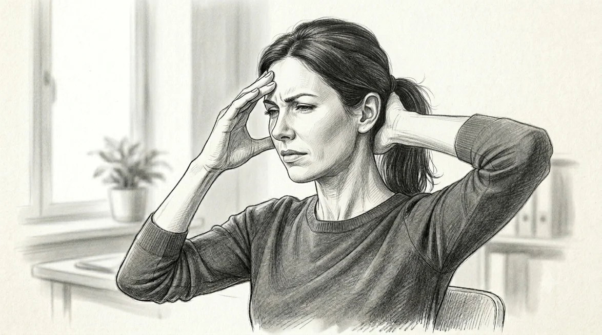
What Is Cervical & Cranial Myofascial Pain?
Myofascial pain refers to pain arising from muscle and its surrounding fascia. Its hallmark feature is the myofascial trigger point — defined as a hypersensitive spot within a taut band of skeletal muscle that, when pressed, produces a familiar referred pain felt at a distance from the spot itself [1]. A trigger point is called active when its local and referred pain reproduce a symptom the person already recognises — for example, the exact headache they get [1].
What makes the neck and head region distinctive is where these muscles refer to. The classic referral maps, documented muscle-by-muscle in Travell & Simons' Trigger Point Manual [2], describe patterns that look a great deal like common headaches:
| Muscle | Common referral zone |
|---|---|
| Sternocleidomastoid (SCM) | Forehead, eyebrow, cheek, ear/mastoid, behind the eye — the clavicular division is also classically associated with postural dizziness and disorientation |
| Upper trapezius | Up the side of the neck, behind the ear, into the temple |
| Splenius capitis | The vertex (top of the head) |
| Splenius cervicis | Through the head toward behind the eye (retro-orbital) |
| Suboccipital muscles | Deep, hard-to-localise pain through the back and side of the head |
| Temporalis | The temple, eyebrow and upper teeth |
In a study of people with chronic tension-type headache, multiple active trigger points were present in exactly these muscles — on average seven per person — and their referred pain reproduced the headache pattern in every participant [1]. Notably, the trigger points in the neck muscles (suboccipital, levator scapulae, splenius) referred pain over larger areas than the head muscles did [1].
Who Typically Experiences This?
Desk workers and screen users
Sustained forward-head postures load the suboccipital, upper trapezius and levator scapulae muscles for hours at a time. In clinical practice we commonly see trigger points develop in exactly these muscles in people who spend long days at a screen, and reduced neck mobility and forward-head posture are frequently found alongside chronic headache [3].
People with long-standing headaches
Many people who present with tension-type headache, and a proportion of those with a musculoskeletal contribution to migraine, also have active trigger points and tenderness in the neck and pericranial muscles [3][1]. The connection is not coincidental — see the fascial lens section below.
People with unexplained "eye," "sinus," or temple symptoms
Because SCM, splenius and temporalis trigger points refer toward the eye, forehead and temple, these presentations are sometimes attributed to the eye or sinuses when the driver is in the neck [2]. The presentation can be remarkably convincing — unilateral pressure behind one eye that has been investigated ophthalmologically and found to be normal is a common story.
People with neck-related dizziness or unsteadiness
The sternocleidomastoid and upper trapezius are among the muscles classically linked to a sense of dizziness, imbalance or disorientation of cervical origin [2][4]. This is an area we assess rather than assume. See our dedicated page on cervicogenic dizziness for the full picture.
People with a history of trauma, stress, or prolonged immobilisation
Physical trauma — a motor vehicle accident, a fall, a whiplash injury — reliably triggers fascial densification and trigger point formation in the affected region and in the compensatory patterns that develop around it. Sustained psychological stress is equally potent: elevated sympathetic tone increases muscle guarding, reduces pain thresholds, and directly affects fascial physiology through its influence on inflammatory mediators and autonomic innervation. A patient whose cervical pain and headaches consistently flare during high-stress periods is not imagining the connection — the biology is real.
The Fascial Lens: Why We See This Differently
Most headache assessments look upward — at the head. We also look down and back, at the cervical muscles and fascia that feed into the same nervous-system junction box as the head and face. But our approach goes further than simply identifying trigger points and treating them in isolation. Understanding why trigger points form and persist in the cervical region requires engaging with the fascial science that the traditional trigger point model does not fully address.
The Trigger Point Model — Its Strengths and Its Limits
The myofascial trigger point model, developed principally by Travell and Simons across several decades, describes trigger points as arising from a sustained depolarisation of motor end plates — a dysfunctional acetylcholine release cycle that creates a local energy crisis in the muscle sarcomere, sensitises local nociceptors, and generates referred pain through central sensitisation mechanisms.
This model has real clinical utility. The referred pain maps that Travell and Simons documented are reproducible, recognisable, and clinically valuable. In the cervical region they are particularly striking: the clavicular division of the SCM, when irritated, causes dizziness and disorientation as well as head pain; splenius cervicis refers pain specifically through the skull to the retro-orbital region; the suboccipital group produces a deep, diffuse head pain that is difficult for patients to localise. These patterns are not coincidences, and they have helped practitioners explain and treat seemingly neurological presentations without unnecessary investigation.
However, the model has limits that matter in practice. High-quality imaging and histological studies have struggled to consistently identify the structural "knot" that the trigger point model proposes. The energy crisis hypothesis, while plausible, has not been definitively confirmed at the histological level. And most importantly from a clinical standpoint, the traditional model places the primary pathology within the muscle belly — which does not fully account for why trigger point therapy so frequently produces temporary relief without lasting resolution when the surrounding fascial environment is not addressed.
This is where Stecco's fascial science begins to complete the picture — and in the cervical region, it does so in a particularly interesting way.
Hyaluronan, Densification and the Loss of Fascial Gliding
Between the layers of the deep fascia — and between the fascia and the underlying muscle — lies a thin layer of loose connective tissue rich in hyaluronic acid (hyaluronan, or HA). In its normal low-viscosity state, hyaluronan acts as a biological lubricant that allows the fascial layers to glide freely over one another as the body moves. When the cervical muscles sustain chronic low-level loading — the kind generated by hours of forward head posture, by the elevated sympathetic tone of psychological stress, or by the guarding that follows a cervical injury — the hyaluronan in the surrounding fascial layers shifts from its normal fluid form to a more viscous, aggregated form. This state — which the Steccos termed densification — reduces or eliminates fascial gliding and significantly increases the stiffness of the fascial compartment.
The muscles can no longer change shape freely. Their force-generating capacity is compromised. And crucially, the mechanoreceptors embedded within the densified fascial layers — Ruffini corpuscles, Pacinian corpuscles, and free nerve endings, all present in the deep cervical fascia in significant densities [5] — receive abnormal mechanical input. Rather than reporting normal gliding and load distribution, they feed a continuous stream of aberrant signals to the central nervous system, which interprets this as a threat, increases local muscle tone, and perpetuates the cycle.
Why this matters clinically
If trigger points are perpetuated by fascial densification — by the inability of fascial layers to glide — then treating the trigger point in isolation (dry needling, ischaemic compression, massage) may produce only temporary relief. The densification remains. The abnormal mechanosensory input continues. The trigger point returns.
This is the mechanism behind the frustrating recurrence that many patients with cervical and cranial myofascial pain experience — and it is precisely why our approach targets the fascial matrix directly, not just the tender spot within it.
The Cervical Fascial System and the Epimysial Argument
Here is an observation that follows from the fascial anatomy but is rarely made explicit in clinical discussions of trigger points.
Cadaveric studies of the cervical fascial system — notably the anatomical work of Natale, Stecco and colleagues (2015) — have documented in detail how the cervical fascia organises itself into multiple investing layers: the superficial cervical fascia, the deep (investing) cervical fascia, the pretracheal fascia, and the prevertebral fascia, all of which are continuous, multi-layered, and richly innervated [5]. Embedded within and continuous with this fascial framework is the epimysial fascia — the dense connective tissue sheath that individually wraps each muscle of the neck.
The sternocleidomastoid has its own epimysial layer that blends directly into the investing cervical fascia. The upper trapezius epimysium merges with the posterior cervical fascia and the thoracolumbar fascia inferiorly. The splenius capitis and cervicis sit within compartments defined by the deep fascial layers, their epimysial sheaths contributing to the compartment wall. Every cervical muscle is enveloped in a fascial sheath that is anatomically continuous with the broader cervical fascial system.
This anatomical continuity invites a question about Travell and Simons' referral maps. The classical model attributes the referred pain patterns to the motor end plate pathology — specifically to the acetylcholine-driven energy crisis cascade. But why would a dysfunctional end plate in the clavicular head of the SCM refer pain specifically to the eyebrow and ear, while one in the sternal head refers to the occiput and vertex? Why would a trigger point in splenius cervicis refer pain specifically through the skull to the retro-orbital region? Why are these patterns so anatomically specific and so reproducible across different patients?
One compelling possibility is that these patterns reflect not only neural sensitisation via the trigeminocervical nucleus, but also the routes of fascial force transmission through the cervical fascial system. A densified epimysial layer within the SCM alters the tension transmitted through the investing cervical fascia toward the mastoid attachment, the fascial connections around the auricular region, and ultimately toward the cranial base. A densified fascial compartment surrounding the splenius cervicis alters the tension path toward the posterior cranium and through the suboccipital fascial continuations. The Travell and Simons referral maps may be mapping, in part, these fascial force-transmission pathways — not only the neurological consequences of end-plate dysfunction.
The two models are not mutually exclusive. The end-plate pathology is real; the neurological sensitisation via the trigeminocervical nucleus is real. But the fascial model explains why the patterns are as anatomically specific as they are in ways the end-plate model alone does not fully account for. And it explains why you cannot reliably resolve these patterns by treating only the muscle belly.
The Trigeminocervical Nucleus — Where Neck and Head Converge
That junction box we mentioned — the trigeminocervical nucleus — is the region of the upper spinal cord where sensory nerves from the upper neck (C1–C3) and the trigeminal nerve (which serves the face and much of the head) converge onto shared neurons [6][7]. Because these inputs share the same neural real estate, a continuous stream of nerve signals from irritated neck muscles can be interpreted by the brain as pain in the head or face. Fernández-de-las-Peñas and colleagues describe precisely this mechanism: active trigger points in muscles innervated by C1–C3 or the trigeminal nerve produce an ongoing afferent barrage into the trigeminal nucleus, which can sensitise the system over time [1].
When the fascial environment of the cervical muscles is densified, the mechanosensory input from the fascial receptors adds to this afferent load. The trigger point generates peripheral sensitisation; the densified fascia generates abnormal mechanosensory input; together they maintain a sustained barrage into the trigeminocervical nucleus that the CNS increasingly interprets as head and face pain. This is how a longstanding neck problem produces what feels entirely like a head problem.
Put simply: the painful spot in your head may be the destination, not the origin. We aim to assess and treat the whole cervical-cranial system rather than only the place it hurts.
The fascial picture — Cervical & Cranial Myofascial Pain
Trigger points are real. The referred pain maps are real. The clinical relief from trigger point therapy is real. But the full picture requires the fascial layer: densified hyaluronan reduces fascial gliding in the cervical compartments, creating the mechanical environment in which trigger points form and persist. The epimysial fascia of every cervical muscle is continuous with the broader cervical fascial system — which means that the specific referral patterns Travell and Simons documented may map both the neurological consequences of end-plate sensitisation and the routes of fascial force transmission through that system.
Treating the trigger point without treating the fascia is like removing the smoke without addressing the fire.
Centres of Coordination — The Stecco FM Model
Stecco's Fascial Manipulation model identifies specific anatomical points within the fascia — called centres of coordination (CCs) — where the vectors of myofascial force for a given movement direction converge. These CCs correspond closely to the trigger point locations documented by Travell and Simons: they are the points where fascial densification is most mechanically significant and where manual intervention produces the greatest change in regional fascial tension.
In the cervical and cranial region, the relevant CCs include points within the investing cervical fascia over the SCM, the posterior cervical fascia over the suboccipital and upper trapezius muscle groups, and points along the lateral cervical fascial chain. A systematic assessment of these points — guided by the movement directions that provoke symptoms — identifies which fascial segments are most dysfunctional and need to be addressed first.
The key distinction between the FM approach and conventional trigger point therapy is not where the hands are placed — it is what the hands are doing and why. In FM, the practitioner applies sustained, specific pressure at the identified CC to generate sufficient local tissue heat to reduce hyaluronan viscosity and restore fascial gliding. The intervention is guided by a systematic biomechanical assessment — not simply by where the patient reports the most pain — because the most painful point in the head is frequently not the primary dysfunctional point in the cervical fascial system. The most painful point is often the destination of fascial force transmission from a more proximal densification.
See also: Myofascial Pain Syndrome — Low Back for the same argument applied to the thoracolumbar fascial system.
What Does the Research Say?
Trigger-point referral reproduces real headaches. In a study of people with chronic tension-type headache, active trigger points in the head, neck and shoulder muscles reproduced the participants' familiar headache pattern, with a mean of seven active points per person [1]. The finding that every participant had their headache reproduced from trigger points is clinically significant — it is not a correlation, it is a mechanical reproduction of the symptom.
Neck muscles carry disproportionate weight. The referred-pain areas from neck-muscle trigger points (suboccipital, levator scapulae, splenius) were significantly larger than those from head muscles, underlining the relevance of the neck in what patients and clinicians may experience as a pure head problem [1].
Cervical impairments are consistently found in headache populations. A large systematic review and meta-analysis (77 studies, 2551 participants) found reduced neck mobility, altered posture, and increased trigger points and tenderness associated with both tension-type headache and the musculoskeletal contribution to migraine — though the authors note the evidence base is still developing and effect sizes are modest [3].
A coherent neurological mechanism exists. Convergence of upper cervical and trigeminal inputs at the trigeminocervical nucleus offers a well-supported explanation for why neck muscle dysfunction can present as head and face symptoms [6][7]. The research on this mechanism has moved from primarily animal studies toward human clinical investigation over the past decade.
The cervical fascia is anatomically distinct and clinically relevant. Cadaveric dissection confirms the deep cervical fascia as a multi-layered, richly innervated structure whose architecture directly influences cervical muscle mechanics and neural element environment — providing the anatomical basis for a fascial approach to cervical myofascial pain [5].
The dizziness link is recognised. The SCM and upper trapezius have a long-documented association with dizziness and disorientation of cervical origin [2][4][7]. In practice, this means a presentation of dizziness and head pain together — with no vestibular findings — is a presentation worth assessing from the cervical myofascial angle.
The literature here ranges from foundational clinical description [2] to modern mechanistic and observational studies [1][6]. The research suggests these patterns are real and assessable — it does not promise that every headache or dizziness has a muscular cause.
How We Approach Cervical & Cranial Myofascial Pain
Our assessment looks beyond the symptom site. We examine the cervical and pericranial muscles and their fascial environment, neck movement and posture, and whether pressing a given muscle reproduces your familiar symptom — the feature that distinguishes a relevant trigger point from an incidentally tender spot [1].
We assess the full cervical fascial system using Stecco FM palpatory protocols — identifying the specific centres of coordination where densification is most significant. This assessment is guided by the movement directions that provoke symptoms, not by the location of maximum pain. The most dysfunctional fascial point is often proximal to — or remote from — the site of maximal discomfort.
Where we identify a loaded, poorly-gliding myofascial environment, our treatment may include:
- Fascial Manipulation (Stecco method) — sustained manual work at the specific cervical and suboccipital centres of coordination, directed at restoring hyaluronan fluidity and fascial gliding
- Manual therapy to the cervical and upper thoracic spine — joint mobilisation and manipulation where segmental hypomobility is contributing to the fascial load
- Trigger point therapy — direct manual pressure or dry needling to address active trigger points once the surrounding fascial environment has been prepared; treating the trigger point without first preparing the fascial matrix is less effective
- Movement and postural retraining — addressing the forward head posture, sustained load patterns, and thoracic mobility restrictions that perpetuate the fascial dysfunction
The goal of treatment is to address the mechanical and fascial environment driving the referred symptoms, not simply to chase the spot that hurts.
New to Fascial Manipulation? Read how it works → · How chiropractic adjustments work →
Please note: The information on this page describes our general clinical approach and is intended for educational purposes only. Individual presentations vary, and your assessment and management will be tailored specifically to you. Nothing on this page constitutes clinical advice for your individual situation. Please consult a registered health practitioner for advice about your specific condition.
What Can You Do Right Now?
1. Reduce sustained loading on the posterior cervical muscles.
Break up long periods of forward-head posture at the screen with regular movement — even brief, frequent changes of position help offload the suboccipital and upper-trapezius muscles. A two-minute movement break every 30–40 minutes is more effective than a longer break once every two hours. Gentle cervical retraction (chin tucks) during these breaks activates the deep cervical flexors and mechanically decompresses the posterior cervical structures.
2. Slow, sustained loading — not rapid stretching.
Rapid stretching does not effectively address fascial densification. The viscoelastic properties of hyaluronan mean it responds to sustained load over time, not to brief high-velocity deformation. A slow, sustained side-flexion or rotation stretch held for 90 seconds — with conscious attention to the gradual release of resistance rather than a sharp pull — is more likely to produce a meaningful change in fascial viscosity than a conventional 20-second stretch performed repeatedly.
3. Address the stress component — it is not in your head.
If your cervical and head symptoms reliably worsen during periods of psychological stress, this is biology, not coincidence. Elevated sympathetic tone directly increases muscle guarding and fascial stiffness. Diaphragmatic breathing is often used to help reduce sympathetic tone, which — through the mechanical effect of diaphragmatic excursion on the cervical fascial system — may in turn ease cervical muscle tension. Sleep quality is a significant perpetuating factor for myofascial pain; it is worth treating as part of the clinical picture.
4. Hydration matters more than you might expect.
Hyaluronan is a highly hydrophilic molecule — it binds water and relies on adequate hydration to maintain its lubricating properties. Chronic mild dehydration directly increases HA viscosity and promotes fascial densification. This is a genuine and underappreciated perpetuating factor, particularly for people who drink mostly coffee and forget water across a working day.
5. Track your triggers.
Noting when symptoms appear — after long desk sessions, specific postures, particular stressors — gives both you and your practitioner useful information about which perpetuating factors are most active. The pattern often clarifies the priority for management more than any single clinical finding.
Related conditions: Cervical Facet Syndrome · Neck Pain in Desk Workers
If you also have any of the following, please seek prompt medical assessment rather than self-managing: a sudden "worst-ever" headache, headache with fever or neck stiffness, new neurological symptoms (weakness, slurred speech, vision loss), or dizziness with these features.
Take the Next Step
Ready to get on top of this?
Call Now — speak with our team
Book Online — available 24/7
Frequently Asked Questions
Further reading
Related articles from the blog
The comprehensive pillar article on the fascial and neurological architecture of the cervical spine — covers the trigeminocervical nucleus, deep cervical fascia, myofascial slings, and how neck dysfunction generates head symptoms across multiple presentations.
Read Article →How the trigeminocervical convergence zone explains why a single fascial and neurological system can produce symptoms that appear to come from three different places at once — and what this means for assessment and treatment.
Read Article →References
- PubMed Fernández-de-las-Peñas C, Ge HY, Alonso-Blanco C, González-Iglesias J, Arendt-Nielsen L (2010). Referred pain areas of active myofascial trigger points in head, neck, and shoulder muscles, in chronic tension type headache. Journal of Bodywork & Movement Therapies, 14(4), 391–396.
- Simons DG, Travell JG, Simons LS (1999). Travell & Simons’ Myofascial Pain and Dysfunction: The Trigger Point Manual, Vol. 1 — Upper Half of Body (2nd ed.). Williams & Wilkins.
- PubMed Pensri C, Liang Z, Treleaven J, Jull G, Thomas L (2025). Cervical musculoskeletal impairments in migraine and tension-type headache: a systematic review and meta-analysis. Musculoskeletal Science and Practice.
- PubMed Li Y, Yang L, Dai C, Peng B (2022). Cervicogenic dizziness: a narrative review of pathogenesis, diagnosis, and treatment. Journal of Clinical Medicine, 11(21), 6293.
- PubMed Natale G, Condino S, Stecco A, Soldani P, Belmonte MM, Gesi M (2015). Is the cervical fascia an anatomical proof for a cervicogenic headache? Surgical and Radiologic Anatomy, 37(4), 385–392.
- PubMed Pankrath F, Bizetti Pelai E, Sobral de Oliveira-Souza AI, Baghbaninaghadehi F, Dennett L, Svensson P, von Piekartz H, Armijo-Olivo S (2025). Nociceptive integration across orofacial, cranial and cervical regions through the trigeminocervical nucleus: a scoping review. Journal of Oral & Facial Pain and Headache.
- PubMed De Hertogh W, Micarelli A, Reid S, Malmström EM, Vereeck L, Alessandrini M (2025). Cervicogenic dizziness — pathophysiology, diagnostic challenges, and therapeutic implications. Frontiers in Neurology, 16, 1545241.
- PubMed Gerwin RD (2001). Classification, epidemiology, and natural history of myofascial pain syndrome. Current Pain and Headache Reports, 5(5), 412–420.