Ready to get on top of this?
📞 Call Now — speak with our team
🗓 Book Online — available 24/7
What Is Cervicogenic Headache?
Cervicogenic headache (CGH) is a secondary headache — meaning its source is not within the head itself, but in the structures of the upper cervical spine, suboccipital region, and associated soft tissues. Pain is referred from these structures to the head via the trigeminocervical nucleus: a region of the brainstem and upper spinal cord where cervical afferent signals from C1, C2, and C3 converge with trigeminal signals from the face and head. When upper cervical joint or soft tissue structures generate sufficient nociceptive input, this convergence creates the subjective experience of headache pain in the head — even though the pain source is in the neck.
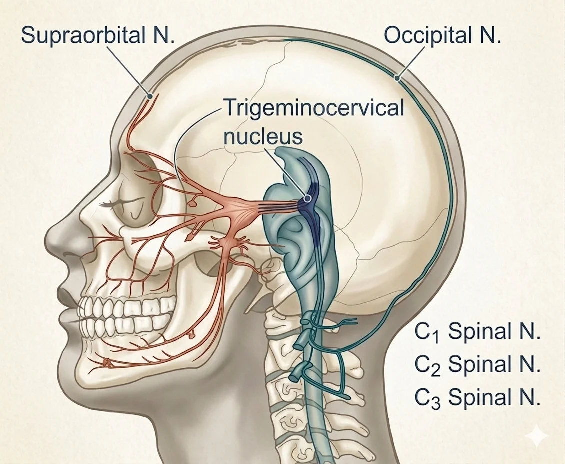
CGH is estimated to account for approximately 15–20% of all chronic headaches. It is frequently confused with tension-type headache and migraine, which delays appropriate treatment. The distinguishing features are its unilateral presentation, its reproduction by neck movement or sustained postures, and its response to manual therapy targeting the upper cervical spine.
Presentation overview
| Feature | Detail |
|---|---|
| Pain location | Typically unilateral; occipital, temporal, frontal, or periorbital — but originating from the neck |
| Symptom pattern | Worsens with sustained neck postures or movement; reduced cervical range of motion; neck stiffness |
| Key diagnostic feature | Pain reproduced by manual pressure on upper cervical joints (C0–C1, C1–C2, C2–C3); diminished by cervical anaesthetic block |
| Differentiation from tension headache | Typically unilateral; associated with neck stiffness and restricted ROM; responsive to cervical manual therapy |
| Differentiation from migraine | CGH lacks the prodrome, photophobia/phonophobia triad, and nausea pattern of migraine — though overlap presentations exist |
| Most affected | Desk workers with sustained cervical flexion posture; people post-whiplash; those with upper cervical joint dysfunction |
Who Typically Experiences This?
Desk workers and screen users
The single most common driver of cervicogenic headache in clinical practice is sustained cervical protraction — the forward head posture of desk work, smartphone use, and prolonged screen time. For every centimetre of forward head translation, the effective weight borne by the cervical spine increases substantially. This sustained loading concentrates at the upper cervical segment and the suboccipital region: the craniocervical junction where C0–C1, C1–C2, and C2–C3 joints are most affected by forward head positioning. The result is cumulative suboccipital and upper cervical joint loading, progressive densification of the suboccipital fascial system, and — eventually — headache referred into the temporal and frontal regions via the trigeminocervical pathway.
People post-whiplash
Acceleration-deceleration injury produces multi-tissue loading across the cervical spine, with the upper cervical segment often receiving significant compressive and shear force. Chronic post-whiplash presentations frequently include a cervicogenic headache component that persists well beyond the acute healing phase. This reflects ongoing mechanical dysfunction at the upper cervical joints and densification of the cervical fascial system following the initial insult. The headache is often attributed to the whiplash itself — but it is the persistent upper cervical mechanical dysfunction that sustains it.
Athletes with high cervical load
Contact sport athletes, swimmers with high training volumes, and weightlifters performing heavy overhead and axial loading are exposed to significant cervical joint stress. When upper cervical joint mechanics become restricted — through cumulative loading without adequate recovery — CGH can develop as a presenting complaint even without a single precipitating event.
The person who has tried multiple headache treatments
A significant proportion of people presenting with cervicogenic headache have previously been treated for tension headache or migraine, often unsuccessfully or with partial response. Prophylactic medications, over-the-counter analgesics, and migraine-specific treatments may reduce symptom severity without addressing the cervical mechanical driver. If headaches are reliably associated with neck stiffness, specific postures, or reproducible by cervical pressure — the cervical source deserves specific assessment.
The Fascial Lens: Why We See This Differently
The cervical fascial layers: a complex system under sustained load
The neck is enclosed in multiple fascial layers — the superficial cervical fascia, the investing deep cervical fascia, the pretracheal fascia, the prevertebral fascia, and the visceral fascia — each with specific anatomical relationships and clinical implications [1]. These layers are not independent structures; they form continuous fascial compartments that transmit tension along predictable pathways. When sustained cervical protraction loads the posterior cervical system, these fascial layers are placed under cumulative tensile and compressive stress. The resulting densification — increased hyaluronan viscosity in the loose connective tissue between layers — reduces normal fascial gliding and concentrates mechanical load at specific segments, including the craniocervical junction.
Fascial Manipulation directed at the relevant centres of coordination in the suboccipital region and cervical fascial system aims to restore normal gliding between these layers, reducing the sustained mechanical input to the upper cervical joints.
Deep cervical flexor insufficiency: the structural contribution
The deep cervical flexors — longus colli and longus capitis — are the primary segmental stabilisers of the cervical spine. They maintain the normal cervical lordosis and resist the sustained anterior shear of forward head posture. Research confirms that deep cervical flexor (DCF) activation is impaired in people with chronic neck pain: the superficial flexors (sternocleidomastoid, scalenes) compensate, creating a high-load, low-control movement pattern that increases segmental stress at the upper cervical joints [2]. This DCF insufficiency is not merely a consequence of neck pain — it perpetuates the mechanical loading pattern that drives cervicogenic headache.
Targeted craniocervical flexion exercise (CCFE) specifically restores DCF activation and reduces the superficial muscle overactivity that compresses the upper cervical segment [2]. This exercise modality is a specific and evidence-supported component of cervicogenic headache rehabilitation.
The myodural bridge: a plausible mechanism, not a proven one
A second idea comes up alongside this one. The deepest of the small muscles at the base of the skull connects, through soft tissue, directly to the dura — the membrane surrounding the brain and spinal cord. That the connection exists is well established: a systematic review of 26 anatomical studies found strong evidence for it from three separate suboccipital muscles [3].
What it does is another matter. The proposal runs like this — thickening of that connection stiffens the dura, reduces compliance at the junction between skull and neck, and alters cerebrospinal fluid flow, while also placing traction on the pain-sensitive dura itself; both signals then arrive at the same brainstem relay described above. In an animal model, thickening this connection produced headache-related behaviour and raised pain-signalling molecules in the brainstem [4].
In people, the picture is less tidy. Imaging studies disagree about whether the muscle is altered in headache at all — it has been reported larger in one chronic headache group, smaller in tension-type headache, smaller again with fatty infiltration in cervicogenic headache, and no different from healthy controls in migraine [413, 414]. To date, this causative link has yet to be demonstrated in humans, and is currently considered a plausible but unproven contributor to headache.
As it stands, this is an interesting anatomical picture whose significance is unresolved. We will be watching this space, and updating this page as new information comes to light.
What Does the Research Say?
Manual therapy is effective for cervicogenic headache — both manipulation and mobilisation
A systematic review of manual therapies for cervicogenic headache assessed 8 RCTs and found that spinal manipulation and mobilisation produce significant reductions in headache frequency, intensity, and disability [5]. Effect sizes are comparable to prophylactic medication. The review supports multimodal manual therapy approaches — combining cervical joint work with soft tissue techniques — as superior to unimodal intervention.
Conservative manual therapy and exercise produce clinically meaningful outcomes
A systematic review of conservative physical therapy management for cervicogenic headache found that manipulation, mobilisation, and exercise each demonstrate benefit [6]. Combining manual therapy with targeted exercise — particularly deep cervical flexor training — produces superior outcomes compared to either treatment in isolation. Deep cervical flexor strengthening is the most consistently supported exercise modality across the included trials.
The suboccipital muscles are physically connected to the dura — what that means clinically is unresolved
A systematic review of 26 anatomical studies found strong evidence that three of the suboccipital muscles connect through soft tissue to the cervical dura [3]; the original dissection describing this proposed that the connection helps resist inward folding of the dura during neck movement [7]. Whether tension at this site generates headache in people has not been demonstrated, and imaging studies in headache populations disagree with one another [413, 414]. We treat the suboccipital region because of its role in upper cervical afferent input, which is well supported — not on the strength of this proposed mechanism.
Deep cervical flexor exercise specifically restores impaired neuromuscular control
A randomised controlled trial found that craniocervical flexion exercise training significantly improves deep cervical flexor activation, pain, and disability in chronic neck pain — while general neck exercise improves pain and disability but does not restore DCF neuromuscular control [2]. This distinction is clinically important: DCF impairment requires specific targeting, and its restoration is associated with improved segmental control at the upper cervical spine.
Fascial Manipulation — evidence across MSK conditions
A systematic review of fascial manipulation across musculoskeletal conditions found evidence supporting its effectiveness for pain and disability in MSK presentations [8]. Applied to the cervical region, FM assessment of the suboccipital and cervical fascial system targets the densification pattern that sustains the mechanical loading of the upper cervical joints.
How We Approach Cervicogenic Headache
Upper cervical assessment
Our assessment identifies which upper cervical segments are contributing to the headache — using manual joint assessment, craniocervical flexion testing, and reproduction of the headache with cervical palpation as the primary clinical tools. We assess for restricted range of motion, segmental joint stiffness at C0–C2, and the postural and movement patterns that load the upper cervical system.
Fascial Manipulation assessment
We assess the suboccipital region — RCPM, obliquus capitis, posterior atlanto-occipital membrane, and the deep cervical fascial system — for centres of coordination where densification is contributing to restricted craniocervical gliding and to altered afferent input from the region. Using the Stecco FM approach, treatment aims to restore normal fascial gliding in the suboccipital and upper cervical fascial system [8].
Cervical joint mobilisation and manipulation
Consistent with the evidence base, we use joint mobilisation and manipulation at the identified dysfunctional upper cervical segments to restore normal joint mechanics, reduce nociceptive input to the trigeminocervical nucleus, and improve cervical range of motion [141, 142].
Deep cervical flexor rehabilitation
We implement a craniocervical flexion exercise programme to address the DCF insufficiency that perpetuates upper cervical loading. Beginning with low-load activation training and progressing toward functional cervical stability, this component is directed at the neuromuscular pattern that maintains the condition between sessions [2].
New to Fascial Manipulation? Read how it works → · How chiropractic adjustments work →
Please note: The information on this page describes our general clinical approach and is intended for educational purposes only. Individual presentations vary, and your assessment and management will be tailored specifically to you. Nothing on this page constitutes clinical advice for your individual situation. Please consult a registered health practitioner for advice about your specific condition.
What Can You Do Right Now?
1. Assess your screen and sitting posture
The most common driver of cervicogenic headache is sustained forward head posture. Check whether your screen is at eye level, your chair supports a neutral lumbar curve, and your head is not consistently protracted when working. Minor positional adjustments — raising the screen, using a headrest, setting a timer to reset posture every 30 minutes — reduce the cumulative load on the upper cervical system between treatment sessions.
2. Begin chin tuck exercises (craniocervical retraction)
A chin tuck performed gently — drawing the chin back and slightly down without tilting the head, feeling the upper cervical spine lengthen — directly targets the deep cervical flexors and reduces the forward head loading position. Hold for 5–10 seconds, repeat 10 times, two to three sets per day. This is a low-load entry point for DCF activation.
3. Monitor whether your headache is consistently triggered by posture or neck movement
Keep a brief headache diary for one week: note the time of onset, the position you were in beforehand, whether neck movement changes the headache, and whether neck stiffness precedes or accompanies the headache. This information directly informs the clinical assessment and helps distinguish cervicogenic headache from other headache types.
4. Avoid sustained end-range cervical positions
Sustained end-range flexion (looking down at a phone), extension (sleeping without cervical support), or rotation (working with the screen to the side) loads the upper cervical segment at its mechanical limit for extended periods. Adjusting the environment to keep the cervical spine closer to neutral — particularly during the activities that consistently precede headache — is a practical, immediate load management step.
Take the Next Step
Ready to get on top of this?
📞 Call Now — speak with our team
🗓 Book Online — available 24/7
Frequently Asked Questions
Further reading
Related articles from the blog
Cervicogenic headache arises from upper cervical structures that converge at the trigeminocervical nucleus — the same region where neck pain, dizziness and referred head pain share a common pathway. This pillar article maps the full cervical fascial and neurological context.
Read Article →The brainstem receives convergent input from the trigeminal nerve and the upper cervical spine — creating the anatomical basis for headache referred from the neck, jaw and craniocervical region. This article explains how these connections produce the overlapping symptoms seen in CGH.
Read Article →Trigger points in the suboccipital, SCM, upper trapezius and splenius muscles refer pain directly into the head and face through the same trigeminocervical pathway — and frequently co-exist with cervicogenic headache. Covers the fascial mechanism and why these trigger points keep recurring.
Read More →References
- PubMed Natale G, Condino S, Stecco A, Soldani P, Belmonte MM, Gesi M (2015). Is the cervical fascia an anatomical proteus? Surgical and Radiologic Anatomy, 37(9), 1119–1127.
- PubMed Jull GA, Falla D, Vicenzino B, Hodges PW (2009). The effect of therapeutic exercise on activation of the deep cervical flexor muscles in people with chronic neck pain. Manual Therapy, 14(6), 696–701.
- PubMed Palomeque-del-Cerro L, Arráez-Aybar LA, Rodríguez-Blanco C, Guzmán-García R, Menendez-Aparicio M, Oliva-Pascual-Vaca Á (2017). A systematic review of the soft-tissue connections between neck muscles and dura mater: the myodural bridge. Spine, 42(1), 49–54.
- PubMed Song X, Yu SB, Yuan XY, Shah MAA, Li C, Chi YY, Zheng N, Sui HJ (2024). Evidence for chronic headaches induced by pathological changes of myodural bridge complex. Scientific Reports, 14, 5285.
- PubMed Chaibi A, Russell MB (2012). Manual therapies for cervicogenic headache: a systematic review. Journal of Headache and Pain, 13(5), 351–359.
- PubMed Racicki S, Gerwin S, DiClaudio S, Reinmann S, Donaldson M (2013). Conservative physical therapy management for the treatment of cervicogenic headache: a systematic review. Journal of Manual & Manipulative Therapy, 21(2), 113–124.
- PubMed Hack GD, Koritzer RT, Robinson WL, Hallgren RC, Greenman PE (1995). Anatomic relation between the rectus capitis posterior minor muscle and the dura mater. Spine, 20(23), 2484–2486.
- PubMed Arumugam A, Harikesavan K (2021). Effectiveness of fascial manipulation on pain and disability in musculoskeletal conditions: a systematic review. Journal of Bodywork and Movement Therapies, 25, 100–109.
- PubMed Hvedstrup J, Amin FM, Hougaard A, Ashina H, Christensen CE, Larsson HBW, Ashina M, Schytz HW (2020). Volume of the rectus capitis posterior minor muscle in migraine patients: a cross-sectional structural MRI study. The Journal of Headache and Pain, 21, 57.
- PubMed Uthaikhup S, Assapun J, Kothan S, Watcharasaksilp K, Elliott JM (2017). Structural changes of the cervical muscles in elder women with cervicogenic headache. Musculoskeletal Science and Practice, 29, 1–6.