 By Dr Steven Hewitt — Chiropractor · AHPRA: CHI0001115420 · 23 March 2026


The Problem the Research Was Trying to Solve
Practitioners of Fascial Manipulation describe a change in tissue texture under the treating hand as treatment progresses. The Stecco model proposes a specific mechanism: that within the loose connective tissue between fascial layers, a substance called hyaluronan — normally a lubricant that allows adjacent layers to glide freely — can increase in viscosity, becoming more gel-like and restricting movement. This restriction, called densification, is held to be a primary driver of myofascial pain.
The challenge has always been demonstrating this mechanism on imaging. Standard MRI can identify gross pathology — disc herniations, tendon tears, joint effusions — but the loose connective tissue between fascial layers is thin, mobile, and difficult to interrogate with conventional sequences. The densification that FM practitioners palpate as "crunchy" or resistant tissue has been difficult to show on imaging.
Hyaluronan is a glycosaminoglycan — a large molecule with a high affinity for water. The study's reasoning is that in its normal state hyaluronan holds water closely, while in the denser, gel-like state the model describes, more water may sit "free" in the tissue. T1ρ (T1 rho) MRI is sensitive to that difference between bound and unbound water.
The Study: Imaging the Fascia Before and After Treatment
Menon and colleagues at NYU School of Medicine and Johns Hopkins applied T1ρ MRI — a sequence sensitive to glycosaminoglycan content and the ratio of bound to unbound water in connective tissue — to the deep fascia of five people with chronic elbow pain lasting more than three months. [1]
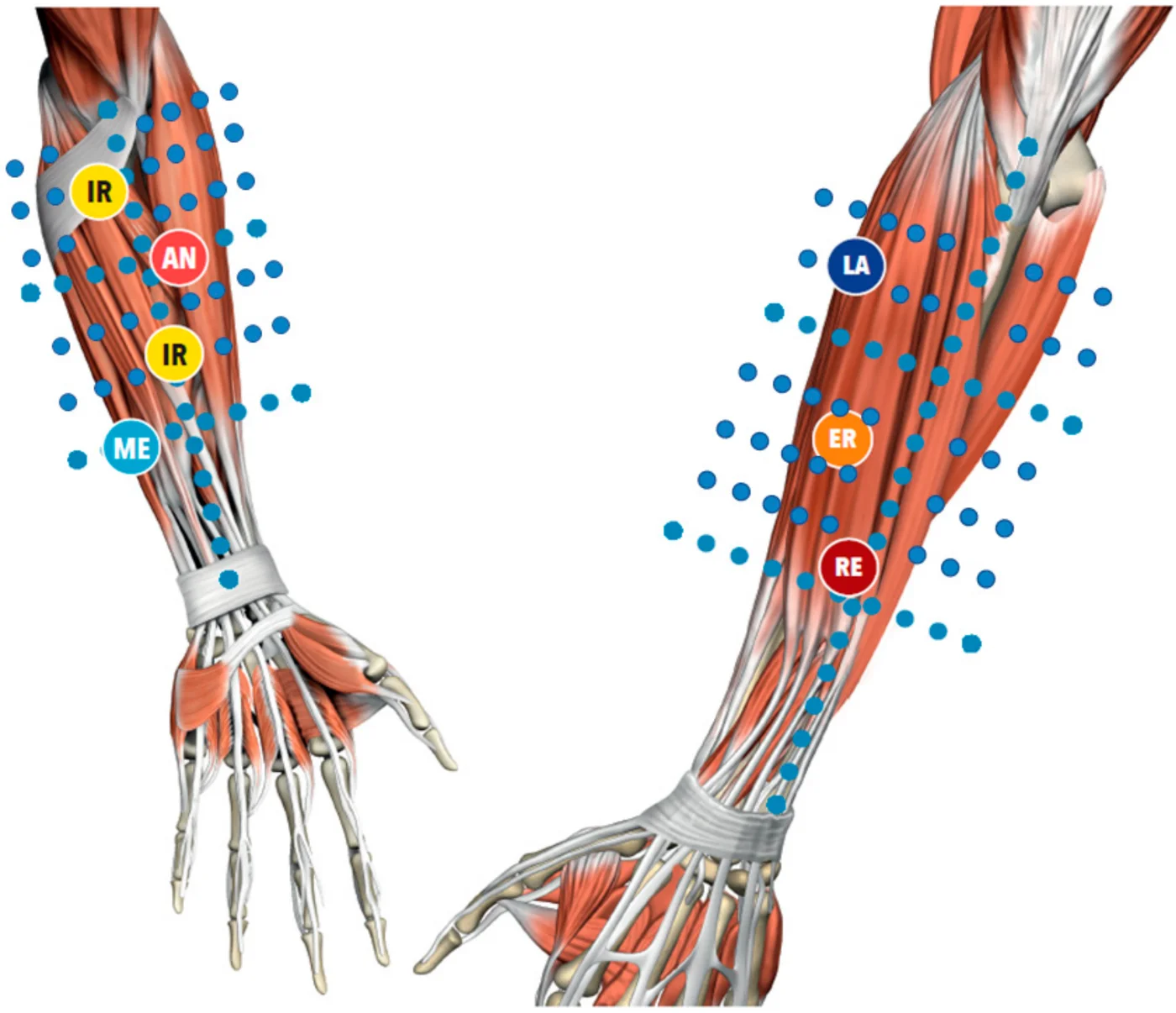
Each received three weekly 40-minute sessions of Fascial Manipulation. T1ρ maps were taken before the first session and after the last; one patient could not be rescanned, so four were imaged both times.
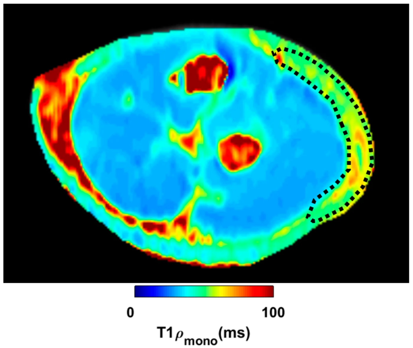
The results went in two directions. In the most severe case, unbound water in the deep fascia fell clearly after treatment, which is the change the densification model predicts. In the other three patients it rose, significantly in two of them. The authors propose the rises could reflect a redistribution of water within the tissue.
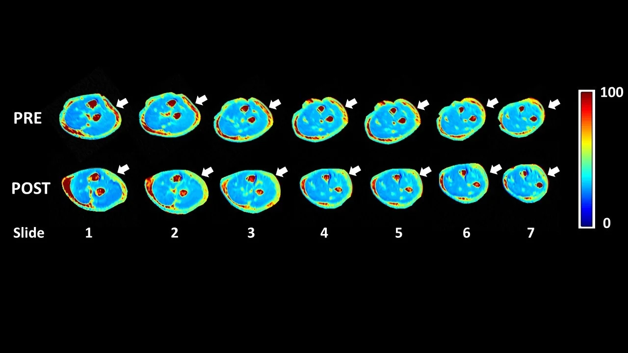
This is not a large trial. It was a case series of five patients, a proof-of-concept design for a new imaging application, with no control group. Its value lies in showing that bound and unbound water in living fascia can be measured before and after treatment, which gives future studies a way to test the densification model properly.
Why This Matters for Our Approach
The densification model is the foundation of Fascial Manipulation. The Stecco method is built on the hypothesis that restricted movement in the loose connective tissue between fascial layers drives altered muscle coordination, concentrated load at tendon insertions, and pain at sites that are often remote from the true source of restriction. The treatment itself applies sustained, deep friction at specific anatomical points — the centres of coordination within each myofascial unit — with the goal of generating sufficient local heat to reduce hyaluronan viscosity and restore normal fascial gliding.
That model has always had strong anatomical support. Stecco's laboratory work on cadaveric and live fascial tissue has documented the presence of hyaluronan, its distribution between fascial layers, and its viscosity changes under different conditions. What has been missing is a way to measure the proposed change in living tissue before and after treatment.
The Menon 2020 paper is a first step towards that. It shows that the property the model is concerned with can be measured in a living forearm, and in its most severe case the measurement moved the way the model predicts. It does not yet show that treatment reliably produces that change, because the other three cases did not.
What This Means for You
If you have chronic musculoskeletal pain that hasn't responded to standard treatment, the fascial environment is worth assessing. The Menon study used patients with chronic elbow pain — a common presentation where the tendon is often treated in isolation while the surrounding fascial system is not addressed. Deep fascia surrounds muscles throughout the body, so the same questions apply well beyond the elbow.
If you've been told "the scan is normal," this research is relevant. Standard MRI is not designed to image the loose connective tissue between fascial layers — the very tissue this study was examining. A normal scan neither rules densification in nor out; standard imaging is not designed to assess it.
If you've wondered what manual therapy is actually doing, this study is an early attempt to find out. It shows the change the Stecco model proposes can be measured; whether treatment reliably produces it is a question for larger, controlled studies.
If you are managing chronic elbow pain specifically — tennis elbow, golfer's elbow, or persistent pain around the elbow that hasn't resolved — the fascial environment of the forearm and elbow region is exactly the tissue imaged in this study. Our approach to both lateral and medial epicondylalgia includes assessment and treatment of the forearm fascial compartments, informed by this evidence base.
Want to know if a fascial assessment is right for you?
Reference
- PubMed Menon RG, Oswald SF, Raghavan P, Regatte RR, Stecco A (2020). T1ρ-Mapping for Musculoskeletal Pain Diagnosis: Case Series of Variation of Water Bound Glycosaminoglycans Quantification before and after Fascial Manipulation® in Subjects with Elbow Pain. International Journal of Environmental Research and Public Health, 17(3), 708.
Please note: This post is intended for educational purposes only and does not constitute clinical advice. Individual presentations vary significantly. Please consult a registered health practitioner for advice about your specific condition.