Ready to get on top of this?
📞 Call Now — speak with our team
🗓 Book Online — available 24/7
What Is Heel Pain?
“Heel pain” describes where you feel something, not what is causing it. Most heel pain is mechanical: it comes from the structures that take load through the heel with every step. The most common single cause is plantar fasciitis, which produces pain under the inner front of the heel, worst with the first steps after rest; it is estimated to affect around one in ten people at some point in their life [1].
The other causes behave differently. Some are found under the heel, some at the back of it, some come from nerves, and a few come from the bone itself or from conditions elsewhere in the body [1], [2]. Screening for those comes first, and the warning signs are in the frequently asked questions below.
Where it hurts
| What you notice | What it often points to | Read more |
|---|---|---|
| Under the inner front of the heel, worst with the first steps in the morning or after sitting, easing as you walk | Plantar fasciitis (plantar fasciopathy) | Plantar fasciitis |
| A deep, bruise-like ache in the middle of the heel pad, worse barefoot on hard floors | Heel fat pad | — |
| Pain that builds with activity after you’ve increased your training or changed to a harder surface, and hurts when the sides of the heel are squeezed | Calcaneal stress fracture (needs imaging) | — |
| Burning, tingling, shooting pain or numbness in the heel or sole | A nerve: at the inner ankle (tarsal tunnel), a small branch under the heel, or a nerve root in the lower back | Sciatica |
| At the back of the heel, where the Achilles attaches, worse in shoes, uphill or first thing | Insertional Achilles tendinopathy, or the bursa and bony bump beside it | Achilles tendinopathy |
| A child or teenager with pain at the back of the heel during sport, often in a growth spurt | Sever’s disease (calcaneal apophysitis), a growth-plate irritation | — |
| Both heels, with morning stiffness, or pain in the back or other joints too | An inflammatory condition (see your GP) | — |
These are first clues, not a diagnosis, and more than one can be present at once.
Who Typically Experiences This?
The runner who added kilometres
A new goal, more sessions, a few more hills, and then heel pain that is worst on the first steps the next morning. Load that rises faster than the tissue can adapt is the most common story behind plantar heel pain, and the timing of the increase is usually the most useful thing in the history. If the pain instead keeps building during runs and the bone is tender to squeeze, a stress fracture needs ruling out before anything else.
The person on their feet all day on hard floors
Retail, hospitality, nursing, warehouse work: hours of standing and walking on concrete, often in flat or worn shoes. Plantar heel pain is common here, and so is the deep central ache of an overloaded heel fat pad, which tends to be worse barefoot and on hard surfaces.
The person whose heel has already been injected
Cortisone can settle plantar heel pain in the short term, which is why it is often offered. It also carries a risk of plantar fascia rupture and of thinning of the heel’s fat pad [1], so a heel that has had several injections is assessed with that in mind.
The child in a growth spurt who plays a lot of sport
Pain at the back of the heel in a child or young teenager, worse with running and jumping, is often Sever’s disease: irritation where the Achilles pulls on the heel’s growth plate [1]. It is a different problem from adult heel pain and is managed around growth and training load rather than as a tendon or fascia problem.
The person whose heel pain comes with tingling
Burning, tingling or numbness points away from the plantar fascia and toward a nerve. That includes nerves at the ankle and under the heel, and also a nerve root in the lower back, which is worth checking even when there is no back pain at all [1].
The Fascial Lens: Why We See This Differently
The heel is one structure, not two
We tend to think of the Achilles tendon and the plantar fascia as separate: one at the back of the heel, one underneath it. Anatomically they are continuous. Dissection and MRI work from Padova found the plantar fascia continuous with the Achilles paratenon (the sheath around the tendon) at the heel bone, with the thickness of one correlating with the other [3]. An independent group in New Zealand, Germany and Japan found aligned bony trabeculae in the back and underside of the heel bone linking the Achilles to the plantar fascia “in a bridge-like manner” [4].
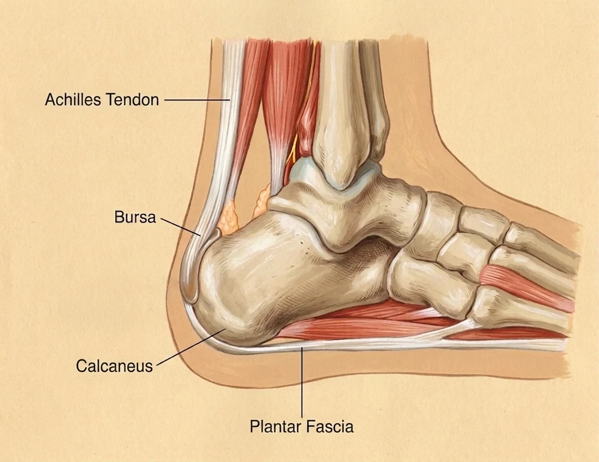
Side view of the heel. The Achilles tendon attaches to the back of the heel bone (calcaneus), with a small bursa beside it; the plantar fascia starts underneath and runs forward towards the toes.
The bone itself seems to record that shared loading. In people with insertional Achilles problems, a spur where the Achilles attaches was accompanied by a spur under the heel far more often than it was absent (65% against 12% in heels without a posterior spur) [5]. The two ends of the heel load together, which is one reason we assess them together.
Upstream, the calf matters
The plantar fascia is the far end of a chain that runs up through the Achilles into the calf. A narrative review from the Pisa group notes that calf and Achilles tightness has been reported in around 80% of people with plantar fasciitis, and that calf-stretching trials have reduced plantar heel pain [6]. The best trials are more mixed than that suggests: a 2021 best-practice review that kept only high-quality trials included one of each, and calf stretching on its own did not beat its comparison, while stretching the plantar fascia itself improved first-step pain in the short term [7]. In people with Achilles tendinopathy, the same group found the fascial plane between the calf muscles more than twice as thick as in matched controls, although the plantar fascia itself was not thicker in that group [8]. A trial that stretched the Achilles and plantar fascia together, rather than the Achilles alone, found a greater improvement in one of seven measures (sensitivity to pressure); its authors hold a patent on the stretching device used [9].
Where you feel it and where it starts
Fascial Manipulation distinguishes the centre of perception, where you feel the pain, from the points of densification that may be changing how load is shared, which often sit in a neighbouring segment. For heel pain those neighbours are the foot, the ankle and the calf, and through the posterior chain, the knee and hip. We look along that line rather than only at the sore spot, and the deep longitudinal sling explains why the back of the leg behaves as one system.
Being straight about the evidence
The anatomical continuity across the heel is well documented by several independent groups. What is not established is that treating the fascia upstream changes heel pain outcomes. The one remote-effect study in healthy people found that treating the plantar fascia changed function further up the chain, but in the direction of slightly less force, not more [10]. We present the fascial lens as a reasoned part of a full assessment, not as a proven treatment for heel pain.
What Does the Research Say?
Most heel pain is mechanical, and location is the best first guide
Clinical reviews organise heel pain by where it is felt: under the heel (plantar fasciitis, fat pad, stress fracture, nerve entrapment), at the back (Achilles insertion, bursa, Sever’s disease in children), and on the inner or outer midfoot (tarsal tunnel, sinus tarsi). Mechanical causes are the most common, and the history and examination usually point to the right one without a scan [1], [2].
A heel spur is linked to heel pain, but hasn’t been shown to cause it
Heel spurs are often blamed for heel pain. They are more common in people with heel pain: a 2009 review pooling seven X-ray studies found spurs much more often in people with chronic heel pain than in people without it, although the studies varied a lot [11]. But spurs also turn up in people with no heel pain at all [1], and no study has shown that the spur itself causes the pain [11]. The same review found the plantar fascia on ultrasound averaged about 2 mm thicker in people with heel pain, again with wide variation between studies [11].
The heel fat pad is a common cause with thin evidence
The fat pad under the heel bone cushions every step. When it is the source of pain, the pain tends to sit in the middle of the heel or around its edge, and to get worse barefoot or after long periods of standing [12]. Two studies suggest it may be the second most common cause of pain under the heel, after plantar fasciitis. In the records of 250 heel pain patients at one Korean clinic, 53% had plantar fasciitis, 15% had fat pad pain and 9% had both; in 784 American adults aged 65 and over, 4.2% had fat pad pain against 6.9% with plantar fascia pain [12].
Beyond that the evidence is thin. A 2022 review found only seven original studies of fat pad pain, mostly small and low on the evidence hierarchy, and no randomised trials of the heel cups and taping usually recommended for it [12]. In the Korean records, pain that worsened with long standing, came on at night or affected both heels made fat pad pain much more likely than plantar fasciitis, while first-step pain and tenderness at the inner front of the heel made it much less likely. Those clues come from one clinic, without adjustment for age or weight, so they are a guide rather than a test [12].
What helps plantar heel pain
A 2021 best-practice guide, which kept only high-quality trials and added the views of experts and patients, put three things at the core of managing plantar heel pain: taping, stretching the plantar fascia, and education tailored to the person, covering footwear, breaking up long periods of standing and how to read pain during activity [7]. For people who don’t improve enough, it suggested shockwave therapy next, and then custom orthoses [7].
Strength training for the heel is less settled. In a trial of 48 people, heavy heel raises with a towel under the toes did better than plantar fascia stretching at three months, but the groups were no different at six and twelve months [13]. A larger trial of 180 people with heel pain lasting three months or more found that adding the same heel-raise programme to advice and a heel cup made no clinically meaningful difference at twelve weeks; all three groups improved meaningfully, including the group given advice and a heel cup alone [14]. The best-practice guide found no high-quality trials of broader strengthening programmes, and the experts it interviewed were divided on them [7].
Manual therapy combined with exercise has been graded as fair evidence for short-term benefit in plantar fasciitis, though on only two studies, with no grade for the long term [15]. Cortisone injections can give short-term relief, with the risks of plantar fascia rupture and fat pad thinning noted above [1]. In the 2023 trial, about twice as many people given an injection on top of the heel raises reached a level of symptoms they were happy with by twelve weeks, but the average difference in pain stayed below what the researchers counted as clinically important, and the differences narrowed over the year [14].
Children’s heel pain is usually a growth-plate problem
Sever’s disease is irritation of the growth plate at the back of the heel in active children and adolescents, and X-rays are usually not needed to diagnose it [1]. Heel raises and orthoses are commonly used for it, but a 2013 review found only nine small studies; the limited evidence favoured orthoses over heel raises for short-term pain relief, and the authors advised caution because of problems with how the studies were designed [16].
How We Approach Heel Pain
Screening first. We check for the features that need a different pathway: a possible stress fracture, signs of an inflammatory condition, nerve symptoms, infection or skin breakdown in people with diabetes, and growth-plate pain in children. If anything points away from a musculoskeletal cause, or needs imaging first, we tell you and arrange it.
Finding where it comes from. Exactly where it hurts, when it hurts, and what provokes it; tests including heel squeeze, first-step and toe-extension provocation, and tapping over the nerves at the inner ankle; and a neurological check that includes the lower back when there is any tingling or numbness.
The chain above the heel. Ankle and big-toe movement, calf and Achilles, and how the knee and hip load the foot when you walk, run or stand on one leg.
The fascial assessment. A Fascial Manipulation assessment across the foot, ankle and calf segments, and further up the posterior chain where the assessment points, looking for densifications that may be changing how load is shared.
Treatment. Typically Fascial Manipulation directed at the areas your assessment identifies as relevant, education about the condition, advice on day-to-day load and footwear, plantar fascia stretching, and a strengthening plan when improving capacity is important. Other approaches, such as taping, shockwave therapy or custom orthoses, may also help; we typically refer out for these when needed, as we do for imaging or a podiatry or medical opinion. Our free Plantar Fasciopathy Rehab Program covers the first two weeks of loading for the most common cause.
New to Fascial Manipulation? Read how it works →
Please note: The information on this page describes our general clinical approach and is intended for educational purposes only. Individual presentations vary, and your assessment and management will be tailored specifically to you. Nothing on this page constitutes clinical advice for your individual situation. Heel pain after a fall or injury, with redness, heat or fever, or with numbness that is spreading, should be assessed medically before a mechanical cause is assumed. Please consult a registered health practitioner for advice about your specific condition.
What Can You Do Right Now?
1. Notice exactly where and when it hurts. Underneath or at the back? Worst on the first steps, or building the longer you’re on your feet? Is there tingling? That pattern is the most useful thing to bring to an assessment.
2. Take the edge off the first steps. Many people find a minute of moving the ankle and toes, or a gentle calf and foot stretch, before getting out of bed makes the first steps easier.
3. Look at your shoes and your floors. A cushioned, supportive shoe usually suits a sore heel better than a flat, worn one or bare feet on hard floors, especially if the pain is in the middle of the heel pad.
4. Ease back on the load spike, not on all movement. If the pain followed a jump in training or time on your feet, bring that back down for a while rather than stopping altogether. Pain that keeps building during activity is a reason to get the bone checked.
5. Know the warning signs. Read the urgent-care question below once, so you would recognise them.
Take the Next Step
Ready to get on top of this?
📞 Call Now — speak with our team
🗓 Book Online — available 24/7
Frequently Asked Questions
Related reading
References
- PubMed Tu P (2018). Heel pain: diagnosis and management. American Family Physician, 97(2), 86–93.
- PubMed Tu P, Bytomski JR (2011). Diagnosis of heel pain. American Family Physician, 84(8), 909–916.
- PubMed Stecco C, Corradin M, Macchi V, Morra A, Porzionato A, Biz C, De Caro R (2013). Plantar fascia anatomy and its relationship with Achilles tendon and paratenon. Journal of Anatomy, 223(6), 665–676.
- PubMed Zwirner J, Zhang M, Ondruschka B, Akita K, Hammer N (2020). An ossifying bridge – on the structural continuity between the Achilles tendon and the plantar fascia. Scientific Reports, 10(1), 14523.
- PubMed Zhu G, Wang Z, Yuan C, Geng X, Zhang C, Huang J, Wang X, Ma X (2020). A radiographic study of biomechanical relationship between the Achilles tendon and plantar fascia. BioMed Research International, 2020, 5319640.
- PubMed Ryskalin L, Morucci G, Soldani P, Gesi M (2024). Do the fasciae of the soleus have a role in plantar fasciitis? Clinical Anatomy, 37(4), 413–424.
- PubMed Morrissey D, Cotchett M, Said J’Bari A, Prior T, Griffiths IB, Rathleff MS, Gulle H, Vicenzino B, Barton CJ (2021). Management of plantar heel pain: a best practice guide informed by a systematic review, expert clinical reasoning and patient values. British Journal of Sports Medicine, 55(19), 1106–1118.
- PubMed Ryskalin L, Fulceri F, Morucci G, Busoni F, Soldani P, Gesi M (2024). Ultrasonographic measurements of gastro-soleus fascia thickness in midportion Achilles tendinopathy: a case-control study. Annals of Anatomy, 256, 152321.
- PubMed Engkananuwat P, Kanlayanaphotporn R, Purepong N (2018). Effectiveness of the simultaneous stretching of the Achilles tendon and plantar fascia in individuals with plantar fasciitis. Foot & Ankle International, 39(1), 75–82.
- PubMed Gabriel A, Konrad A, Roidl A, Queisser J, Schleip R, Horstmann T, Pohl T (2022). Myofascial treatment techniques on the plantar surface influence functional performance in the dorsal kinetic chain. Journal of Sports Science & Medicine, 21(1), 13–22.
- PubMed McMillan AM, Landorf KB, Barrett JT, Menz HB, Bird AR (2009). Diagnostic imaging for chronic plantar heel pain: a systematic review and meta-analysis. Journal of Foot and Ankle Research, 2, 32.
- PubMed Chang AH, Rasmussen SZ, Jensen AE, Sørensen T, Rathleff MS (2022). What do we actually know about a common cause of plantar heel pain? A scoping review of heel fat pad syndrome. Journal of Foot and Ankle Research, 15(1), 60.
- PubMed Rathleff MS, Mølgaard CM, Fredberg U, Kaalund S, Andersen KB, Jensen TT, Aaskov S, Olesen JL (2015). High-load strength training improves outcome in patients with plantar fasciitis: a randomized controlled trial with 12-month follow-up. Scandinavian Journal of Medicine & Science in Sports, 25(3), e292–e300.
- PubMed Riel H, Vicenzino B, Olesen JL, Bach Jensen M, Ehlers LH, Rathleff MS (2023). Does a corticosteroid injection plus exercise or exercise alone add to the effect of patient advice and a heel cup for patients with plantar fasciopathy? A randomised clinical trial. British Journal of Sports Medicine, 57(18), 1180–1186.
- PubMed Brantingham JW, Bonnefin D, Perle SM, Cassa TK, Globe G, Pribicevic M, Hicks M, Korporaal C (2012). Manipulative therapy for lower extremity conditions: update of a literature review. Journal of Manipulative and Physiological Therapeutics, 35(2), 127–166.
- PubMed James AM, Williams CM, Haines TP (2013). Effectiveness of interventions in reducing pain and maintaining physical activity in children and adolescents with calcaneal apophysitis (Sever’s disease): a systematic review. Journal of Foot and Ankle Research, 6(1), 16.