 By Dr Steven Hewitt — Chiropractor · AHPRA: CHI0001115420 · 11 August 2026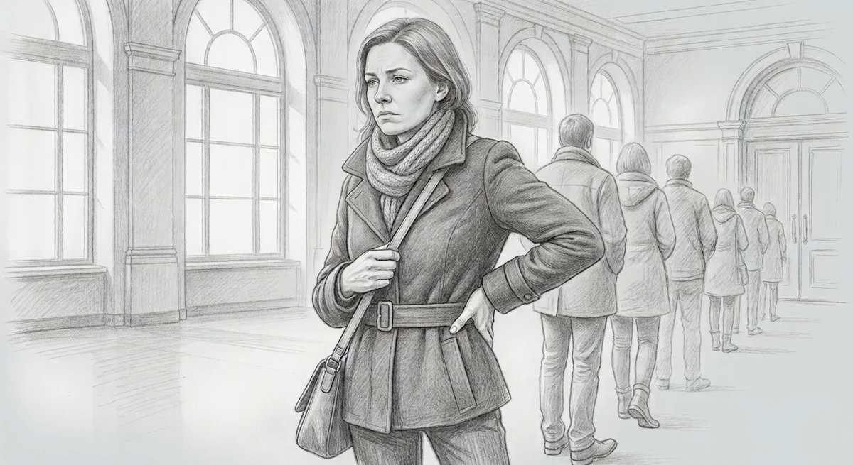
It's one of the most common questions we hear, and one of the most counter-intuitive: "Why does my back hurt when I just stand still — but I can walk for ages and feel fine?" Standing still feels like it should be the easy option. You're not lifting anything, you're not bending, you're barely moving. So why does five minutes in a queue, or a long conversation on your feet, leave your lower back aching when a brisk walk doesn't?
Here's the short answer: for most people, back pain that shows up when you stand still is not a sign of your spine wearing out or being damaged — despite what you may have been told. It's a result of how well, or how poorly, the muscles around your hips and pelvis are managing the job of holding you upright — and there's a surprisingly strong body of research to back that up. There's also one less-common cause worth knowing how to recognise, because it points in a different direction. Let's walk through both.
The 30-second version
- Standing still is a balance task, not a rest task. Holding one position loads the same tissues continuously, with none of the natural on-off contract-and-relax cycling through the muscles that walking gives you.
- The most common driver is poor frontal-plane control at the hip — specifically how your gluteus medius (the muscle on the side of your hip) stabilises your pelvis. Research that asked pain-free people to stand for two hours found that a striking number developed back pain, and the ones who did shared a tell-tale glute pattern before the pain ever started.
- If you've been told it's because of something on a scan — arthritis, a disc bulge, facet changes, a spondylolisthesis, "wear and tear" — the finding is real, but it may not be the reason you can't stand still. Findings like these turn up routinely on the scans of people with no pain whatsoever, and a fixed structural finding that doesn't change from day to day is a poor explanation for pain that comes and goes with what you're doing.
- It's usually very treatable — and often improves with movement strategy and targeted loading rather than rest.
- One pattern is different and worth flagging: if the pain is more in your legs than your back, comes on with walking and standing, and eases the moment you sit down or lean forward over a trolley — that can point to lumbar spinal stenosis, which is worth a proper assessment, especially over the age of 60.
Why standing still is harder on your back than moving
Most people assume the spine "rests" when you stand still. It doesn't. Staying upright is an active, continuous balancing act — dozens of small muscles around the spine, pelvis and hips making constant micro-adjustments to keep your centre of mass — essentially your pelvis — over your base of support, which is your feet.
When you walk, that work is shared and rotated. Each leg takes turns; muscles contract and relax rhythmically; pressure shifts continuously through different tissues. When you stand still, none of that happens. The same structures hold the same position under the same load, minute after minute. Tissues that are perfectly happy being loaded and then released may start to complain when they're loaded and held.
This is why, for a lot of people, walking genuinely feels better than standing. And while standing still looks like resting compared to walking, for the tissues that keep you upright it's closer to the opposite: without the rhythmical contract-and-relax cycles that walking provides, the load never lets up. It simply accumulates — quiet, constant and fatiguing — on the same structures the entire time you're on the spot.
The most common cause: how you control your pelvis, not what your scan shows
This is where the research gets genuinely interesting, because it's been studied directly.
Over a series of studies, researchers took pain-free volunteers — people with no history of back trouble — and simply asked them to stand and do light tasks for two hours. Consistently, a large proportion (in some studies around half, in others up to two-thirds) developed low back pain during that two hours.[1][2] These weren't injured people, and nothing was done to them. Standing still for long enough was all it took.
The crucial finding was what separated the people who developed pain from the people who didn't. It wasn't strength. It wasn't fitness. It was a specific pattern in how their hip and trunk muscles behaved. The people who went on to ache showed co-contraction of the gluteus medius — the muscles on both sides of the hips bracing at the same time, instead of taking turns to control the pelvis side-to-side.[1] In the original study, this glute pattern alone correctly classified most of who would develop pain.[1] A companion study added a second piece: the people who developed pain also had lower endurance in their side muscles — but, tellingly, not lower strength.[2]
Two further details make this more than a laboratory curiosity:
- The pattern comes first. When researchers looked closely, the altered muscle behaviour was present before pain developed — it wasn't a reaction to pain, it was a set-up for it.[3] In laboratory recordings, people prone to standing pain also tend to show a different order of muscle activation when straightening up from a bent-over position — their lower-back muscles switching on before their glutes, where more resilient standers tend to fire glutes first.[4] (That's a pattern measured with electrodes in a lab, not something judged reliably by eye — but it points the same way: the glutes aren't taking their share at the right moment.)
- It predicts the future. People who developed that transient pain during standing were roughly three times more likely to go on to develop genuine clinical back pain over the following couple of years.[5] In other words, an achy back when standing isn't trivial — it's an early signal worth acting on.
And reassuringly, the symptoms produced in these standing studies have been formally checked against the real thing: the quality and location of the pain people get in the two-hour standing test matches the pain of people with everyday clinical low back pain.[6] This is a real-world pattern, not an artefact.
One thing worth saying before we go further, because it’s easy to draw the wrong conclusion from all that. The glute pattern is a good predictor of who develops standing pain — but it doesn’t appear to be the thing you have to fix in order to feel better. When people with standing pain did six weeks of home exercise, how long they could stand improved measurably, while that side-to-side glute pattern stayed much as it was.[14] The goal isn’t to erase a pattern you may have had for years. It’s to widen what your body comfortably tolerates.
If you want the deeper dive on why "lazy glutes" is the wrong way to think about this — and what's actually going on in the muscle — we've written a whole article on it: "Lazy Glutes" Is the Wrong Diagnosis.
"But I was told it's because of what showed up on my scan"
This is worth addressing directly, because it is one of the most common things we hear. Someone can't stand still without their back aching, they've had a scan, the scan found something — and the pain has been attributed to it. From that point on, the finding becomes the explanation for everything, and often for good.
The label varies enormously, and that's rather the point. Arthritis. Degeneration. Wear and tear. A disc bulge, protrusion or "slipped disc". Facet joint arthritis or facet hypertrophy. A spondylolisthesis. An "unstable" segment. Loss of disc height. Bone spurs. Scoliosis. In practice, whatever the report happens to mention tends to become the explanation offered — and if a scan lists several findings, as most do past a certain age, one of them usually gets nominated as the chief culprit. People arrive having been told something along the lines of: "Well, of course your back hurts when you stand for long periods — your scan shows arthritis."
Your scan isn't wrong, and neither is your report. If it says you have facet arthritis or a spondylolisthesis, you almost certainly do — these are careful descriptions by people trained to make them. But a scan can only answer one question: what does this spine look like? It cannot answer the question you actually came in with: why does it hurt after ten minutes on my feet, and not when I'm walking? Those are two separate questions, and it's easy — for patients and clinicians alike — for a confident answer to the first to be taken as an answer to the second.
Here's the context a report can't give you. Researchers pooled the scans of 3,110 people with no back pain at all and worked out how common each finding was at each age. The findings turned out to be the norm rather than the exception:[17]
| Finding on the scan | Pain-free at 20 | Pain-free at 50 | Pain-free at 80 |
|---|---|---|---|
| Disc degeneration | 37% | 80% | 96% |
| Disc bulge | 30% | 60% | 84% |
| Facet joint arthritis | 4% | 32% | 83% |
| Spondylolisthesis | 3% | 14% | 50% |
Not one of those people had any back pain. Their scans simply hadn't been taken — and if they had been, they'd have been handed the same labels, and quite possibly the same explanation. The authors of that study concluded that these changes are "generally part of the normal aging process rather than pathologic processes requiring intervention."[17] A separate study that scanned a community group for an unrelated reason found facet joint arthritis in two out of three people, most of whom had no back pain at all.[18]
None of which means these findings are meaningless, and we won't tell you they are. Ask the sharper question — are they commoner in people who do have back pain? — and for several of them the answer is yes.[19] The point is narrower than "your scan doesn't matter." It's that these findings are common enough that having one doesn't establish it's the thing making you sore.
The clue is usually in how your symptoms behave, not in what your scan shows. Every finding on that list is essentially fixed on the timescale of your day — arthritis doesn't develop between breakfast and lunch, a disc doesn't bulge further while you're queueing, and a vertebra's position doesn't change between Tuesday and Wednesday. That makes any of them a poor candidate for explaining pain that comes and goes with what you happen to be doing. We commonly see people who can get through a full game of sport, or a long walk, completely comfortably — and then ache within minutes of standing still. The spine is identical in both situations. What differs is how long one position is held, and how evenly the load is being shared while it's held. That's the part that responds to being worked on.
Sometimes a finding is genuinely part of the picture. Sometimes it changes what's sensible to do — a large slip is a different situation from a small one — and occasionally, as with the stenosis pattern below, it matters a great deal. Working out which of those applies to you is what an assessment is for, and it isn't something to settle from an imaging report alone. That's the position the research takes on stenosis too: the strongest reviews are clear that imaging on its own isn't enough to make the call, precisely because these findings show up so often in people with no symptoms.[8][9]
The reason we're raising it is simpler than the science. A structural label lands as permanent. It tends to end the conversation, and with it the sense that anything can be done — people stop lifting, stop running, stop standing at their kid's netball game, and quietly accept an aching back as something they have to deal with for the rest of their life. If the actual driver is how load is being distributed while you stand, that's not permanent at all. It's a pattern, and patterns can be assessed, retrained and changed. That's a very different prospect from being told you have a bad back.
(We set out this evidence in more detail on our Lumbar Disc Problems page, and cover the facet joints — what they do, when they genuinely are involved, and how that's worked out — on Lumbar Facet Syndrome.)
The fascial lens: why we look beyond the muscle that's firing
Naming the gluteus medius is only half the picture. The more useful question is why a perfectly capable muscle starts bracing inefficiently when you stand — and this is where our approach differs from a purely "strengthen your glutes" prescription.
Standing balance in the side-to-side direction isn't the job of one muscle. It's the job of a coordinated system — the gluteus medius on one side working with the deep trunk muscles and the opposite quadratus lumborum, all connected through the thoracolumbar fascia, the broad sheet of connective tissue across your lower back that ties your pelvis, spine and hip muscles into one functional unit.[7] When that system shares load smoothly, standing is effortless. When the coordination breaks down — when muscles brace together instead of trading off — load stops being distributed and starts concentrating, and the fascia and muscles that are stuck holding the position fatigue and become sensitised.
This is why we assess standing-related back pain as a load-distribution problem across that whole sling system, not just a weak link to be strengthened in isolation. The muscle that's aching is often not the one that needs the most attention — it's the one left carrying the load when the system upstream or downstream isn't pulling its weight. We explore this network in detail in our piece on the lateral raphe and lumbar load transfer.
There's a postural piece to this, too. How your pelvis sits when you stand — and how deep the curve in your lower back is (the lumbar lordosis) — changes how efficiently these stabilising muscles can do their job. Pelvis position measurably changes how well the glutes fire: in one laboratory study of a side-lying hip exercise, gluteus medius and gluteus maximus were significantly more active with the pelvis held neutral than rolled backwards.[15] And in people prone to standing back pain, a deeper standing curve tends to go hand in hand with more of that inefficient co-bracing of the hip muscles — the two rise together, and they track with the pain. The encouraging flip side: simple adjustments that gently ease that curve — resting a foot up on a step, using a footrest — have been shown to reduce both the co-contraction and the discomfort.[16] It's a good example of why position and load-sharing, not just muscle strength, sit at the centre of how we think about this.
It also reframes the goal. The research is clear that strength wasn't the difference between people who ached and people who didn't — endurance and coordination were.[2][3] So the aim of treatment isn't simply to make a muscle bigger. It's to restore the timing, the endurance and the smooth sharing of load through the whole system — so that holding yourself upright stops being a job a few overworked tissues have to grind through.
When it's not just muscles: spinal stenosis, and how it's different
Most standing-still back pain is the mechanical, very-treatable pattern above. But there's one cause that's worth knowing how to recognise, because it behaves differently and points toward a different kind of assessment: lumbar spinal stenosis.
Stenosis simply means a narrowing of a passageway in the body. Spinal stenosis most commonly refers to a narrowing of the spinal canal centrally — the main channel the nerves run through — which tends to affect the nerves heading into both legs. It becomes increasingly common with age, particularly after 60; the clinical syndrome affects an estimated 11% of adults.[8]
That central narrowing isn't fixed — it changes with the position of your spine. It's accentuated when you stand upright, and especially when you arch backwards (extension): think standing on a downhill slope, or walking downhill, where your lower back has to extend a little to keep you balanced. The same narrowing is relieved by bending forward (flexion), which opens the canal back up. This is why people with central canal stenosis often notice a curious giveaway — walking uphill feels easier than walking downhill. Uphill gently rounds the lower back and reduces the arch (decreasing the lumbar lordosis), opening the canal; downhill increases the arch and narrows it further. When the canal is crowded in those positions, the result is a pattern called neurogenic claudication: pain, heaviness, cramping or pins-and-needles spreading into the buttocks and legs.
Here's the part that trips people up. You might expect a neat split — "muscle pain when standing, nerve pain when walking." It isn't that clean. Stenosis can be aggravated by standing and walking, particularly with the back slightly arched. The genuinely useful distinctions are these:[8][9][10]
| Mechanical / gluteal standing pain (most common) | Lumbar spinal stenosis (worth assessing) | |
|---|---|---|
| Typical age | Any age — very common in working-age adults | Usually older; risk climbs after ~60 |
| Where you feel it | Across the low back / belt line — back-dominant | Often leg-dominant — buttock, thigh, calf; frequently both legs |
| Nerve symptoms | Usually none | Heaviness, pins-and-needles, or leg fatigue/weakness |
| What eases it | Walking, shifting weight, moving around | Sitting and bending forward — the classic "leaning on the shopping trolley" relief |
| What provokes it | Sustained static standing | Standing and walking, especially with the back extended |
That forward-bend relief is the single most telling clue. In a large review of the evidence, the findings that most strongly raised the likelihood of stenosis were no pain when seated, relief when bending forward, and pain in both buttocks or legs — while imaging on its own was not enough to make the call, because narrowing shows up on scans of plenty of people who have no symptoms at all.[8] That last point matters: stenosis is diagnosed from the story and the examination, confirmed by imaging — not from a scan in isolation.
None of this is something to self-diagnose from a table. The point is simply to know when the pattern is worth a conversation with a practitioner.
A quick safety note — when to get it checked sooner
Most standing back pain is mechanical and settles with the right approach. But please see a registered health practitioner promptly if your back pain is accompanied by any of the following:
- Progressive leg weakness, numbness, or pins-and-needles, or any change in bladder or bowel control (this warrants urgent assessment).
- Leg-dominant pain that reliably comes on with walking or standing and eases when you sit or lean forward — especially if you're over 60.
- Back pain that is worse with rest and better with movement, with morning stiffness lasting more than 30 minutes, particularly if you're under 40 — a pattern worth ruling out a different (inflammatory) cause for.
- Pain following a significant fall or injury, or alongside unexplained weight loss, fevers, or feeling generally unwell.
What you can do right now
If your back aches when you stand still, a few evidence-informed strategies are worth trying — and notice that almost none of them is "rest more."
- Trade static standing for micro-movement. The research is unambiguous that holding a position is the problem. Shift your weight, rock gently heel-to-toe, take a few steps, change your stance. Movement redistributes the load the spine has been holding in one place.
- Rest one foot up on a step. A simple, time-honoured trick: prop one foot up on a low step, stool, rail or footrest, and swap feet every minute or two. Bringing one hip and knee up gently flattens the arch in your lower back and takes pressure off the structures that fatigue when you stand — in studies of people prone to standing pain, easing that lower-back curve this way measurably reduced both the muscle co-contraction and the discomfort.[16] (It's why bars have always had a foot rail.)
- Use the forward-bend break. In one study, people who interrupted prolonged standing with brief trunk-flexion breaks — simply bending forward for a few moments periodically — reduced their standing back pain by 36%.[11] The bending momentarily "switches off" and rests the very muscles that fatigue during standing.[11] A short, gentle forward bend every 20–30 minutes on your feet is a genuinely evidence-backed micro-break.
- Keep gently shifting your weight. Rather than standing planted and still, let your weight drift gently onto one foot, then back onto the other, every so often. There's research behind this one: when people are watched during prolonged standing, the ones who don't go on to develop back pain tend to be those who shift their weight and make small movements more — right from the first few minutes, well before any pain appears — while those who stay statically planted are the ones who tend to ache.[13] Like the foot-on-a-step trick, it keeps the load circulating instead of letting it settle into the same tissues — small, unhurried shifts, not constant fidgeting.
- Don't lock out. Standing rigidly with knees braced and back arched concentrates load. A soft, "tall but relaxed" stance breaks up the sustained loading, and anti-fatigue matting helps on hard floors.
- Build hip endurance, not just hip strength. Because the difference between achy and resilient standers is endurance and coordination, the most useful work is generally endurance-based control of the hip and trunk — not heavy isolated glute exercises. Targeted hip-abductor work has good evidence behind it for related hip-control problems,[12] but how you train matters as much as how hard. This is exactly the kind of thing worth getting assessed rather than guessing at. And there's reason for optimism: in one study, people who struggled with prolonged standing followed a progressive home-exercise program for six weeks and significantly improved how long they could stand before pain set in.[14]
And one thing that probably won't help: stretching your hamstrings. The curve of your lower back genuinely does matter here — as above, a deeper standing curve tends to travel with more of that inefficient co-bracing, and gently easing the curve eases the pain along with it.[16] But hamstring length turns out not to be the lever that moves that curve. When 55 adults completed a hamstring stretching protocol, their hamstring length improved measurably — and their standing posture did not change at all. The curve of the lower back, the tilt of the pelvis and the curve of the upper back were all unchanged, by margins smaller than the measuring equipment could reliably detect.[20] The only position that changed was bending forward with the knees straight — the one position that pulls directly on the hamstrings.
These are general, educational suggestions — not a substitute for an individual assessment, particularly if your pain is persistent or follows the leg-dominant pattern described above.
How we approach standing-related back pain
In clinical practice, we treat "my back hurts when I stand" as a question about how you distribute load, not just which structure hurts. Our assessment looks at how your pelvis is controlled both side-to-side and front-to-back, how your hip and trunk muscles share the work of standing, the endurance and timing of that system, and the quality of the fascial tissue that ties it together — rather than assuming the problem lives wherever you happen to feel it. Both directions matter, and the research suggests each does a different job.
From there, treatment is directed at restoring smooth load transfer through that system — combining hands-on Fascial Manipulation to address restrictions in the connective tissue with movement and loading strategies aimed at improving the endurance and coordination of how you hold yourself upright. The goal is to make standing feel like the easy, low-effort task it's supposed to be. And where the pattern suggests something that needs further investigation — like the stenosis picture above — the right first step is a careful assessment, not a guess.
Wondering why your back aches when you're on your feet?
Call Now — speak with our team · Book Online — available 24/7
Prefer to read more first? Our lower back pain page sets out the conditions we commonly assess, and the Lower Back Reset is a free five-module program you can start today.
References
- PubMed Nelson-Wong E, Gregory DE, Winter DA, Callaghan JP (2008). Gluteus medius muscle activation patterns as a predictor of low back pain during standing. Clinical Biomechanics, 23(5), 545–553.
- PubMed Marshall PW, Patel H, Callaghan JP (2011). Gluteus medius strength, endurance, and co-activation in the development of low back pain during prolonged standing. Human Movement Science, 30(1), 63–73.
- PubMed Nelson-Wong E, Callaghan JP (2010). Is muscle co-activation a predisposing factor for low back pain development during standing? A multifactorial approach for early identification of at-risk individuals. Journal of Electromyography and Kinesiology, 20(2), 256–263.
- PubMed Nelson-Wong E, Alex B, Csepe D, Lancaster D, Callaghan JP (2012). Altered muscle recruitment during extension from trunk flexion in low back pain developers. Clinical Biomechanics, 27(10), 994–998.
- PubMed Nelson-Wong E, Callaghan JP (2014). Transient low back pain development during standing predicts future clinical low back pain in previously asymptomatic individuals. Spine, 39(6), E379–E383.
- PubMed Sorensen CJ, Johnson MB, Callaghan JP, George SZ, Van Dillen LR (2015). Validity of a paradigm for low back pain symptom development during prolonged standing. Clinical Journal of Pain, 31(7), 652–659.
- PubMed Willard FH, Vleeming A, Schuenke MD, Danneels L, Schleip R (2012). The thoracolumbar fascia: anatomy, function and clinical considerations. Journal of Anatomy, 221(6), 507–536.
- PubMed Katz JN, Zimmerman ZE, Mass H, Makhni MC (2022). Diagnosis and management of lumbar spinal stenosis: a review. JAMA, 327(17), 1688–1699.
- PubMed Suri P, Rainville J, Kalichman L, Katz JN (2010). Does this older adult with lower extremity pain have the clinical syndrome of lumbar spinal stenosis? JAMA, 304(23), 2628–2636.
- PubMed Genevay S, Courvoisier DS, Konstantinou K, Kovacs FM, Marty M, Rainville J, Norberg M, Kaux JF, Cha TD, Katz JN, Atlas SJ (2018). Clinical classification criteria for neurogenic claudication caused by lumbar spinal stenosis. The N-CLASS criteria. The Spine Journal, 18(6), 941–947.
- PubMed Stewart DM, Gregory DE (2016). The use of intermittent trunk flexion to alleviate low back pain during prolonged standing. Journal of Electromyography and Kinesiology, 27, 46–51.
- PubMed Khayambashi K, Mohammadkhani Z, Ghaznavi K, Lyle MA, Powers CM (2012). The effects of isolated hip abductor and external rotator muscle strengthening on pain, health status, and hip strength in females with patellofemoral pain. Journal of Orthopaedic & Sports Physical Therapy, 42(1), 22–29.
- PubMed Gallagher KM, Callaghan JP (2015). Early static standing is associated with prolonged standing induced low back pain. Human Movement Science, 44, 111–121.
- PubMed Ingerson E, Renfrow C, Aragon E, Ferger N, Olson B, Sachs A, Nelson-Wong E (2019). Individuals with low back pain improve in standing tolerance and sagittal plane muscle activation following exercise intervention. Journal of Back and Musculoskeletal Rehabilitation, 32(6), 885–895.
- PubMed Willcox EL, Burden AM (2013). The influence of varying hip angle and pelvis position on muscle recruitment patterns of the hip abductor muscles during the clam exercise. Journal of Orthopaedic & Sports Physical Therapy, 43(5), 325–331.
- PubMed Abbasi S, Minoonejad H, Abbasi H, Mousavi SH (2025). The impact of different standing positions on gluteus medius activation and lumbar lordosis in LBP-developers during prolonged standing. PLOS ONE, 20, e0317291.
- PubMed Brinjikji W, Luetmer PH, Comstock B, Bresnahan BW, Chen LE, Deyo RA, Halabi S, Turner JA, Avins AL, James K, Wald JT, Kallmes DF, Jarvik JG (2015). Systematic literature review of imaging features of spinal degeneration in asymptomatic populations. AJNR American Journal of Neuroradiology, 36(4), 811–816.
- PubMed Kalichman L, Kim DH, Li L, Guermazi A, Hunter DJ (2010). Computed tomography-evaluated features of spinal degeneration: prevalence, intercorrelation, and association with self-reported low back pain. The Spine Journal, 10(3), 200–208.
- PubMed Brinjikji W, Diehn FE, Jarvik JG, Carr CM, Kallmes DF, Murad MH, Luetmer PH (2015). MRI findings of disc degeneration are more prevalent in adults with low back pain than in asymptomatic controls: a systematic review and meta-analysis. AJNR American Journal of Neuroradiology, 36(12), 2394–2399.
- PubMed López-Miñarro PA, Muyor JM, Belmonte F, Alacid F (2012). Acute effects of hamstring stretching on sagittal spinal curvatures and pelvic tilt. Journal of Human Kinetics, 31, 69–78.
Please note: This post is intended for educational purposes only and does not constitute clinical advice. Individual presentations vary, and your assessment and management will be tailored specifically to you. Nothing here is a diagnosis. Please consult a registered health practitioner for advice about your specific situation.